Characterization of physiological alterations in skeletal muscle by quantitative MRI
(still in progress)
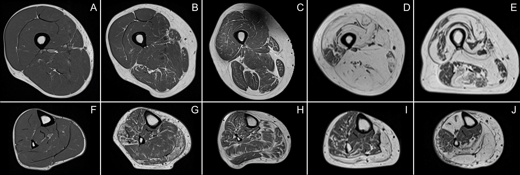
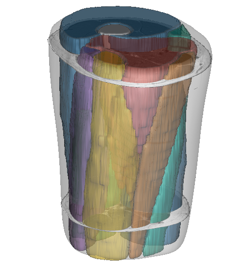
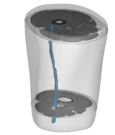
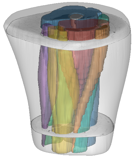
The development of segmentation methods for muscle soft tissues (see the dedicated project here) has allowed the 3D exploration of MRI images on large populations of patients with neuromuscular diseases. The transposition of our methodology into user-friendly tools has enabled us to overcome the barrier of clinical use. Therefore, in collaboration with clinicians from the neurology department of the CHU de la Timone in Marseille, several studies have been conducted on the characterization of physiological alterations occurring at the level of individual muscles in various pathologies and during physical exercises. These studies have notably confirmed the potential of quantitative MRI scores as biomarkers of neuromuscular pathologies.

The pattern of fat infiltration by qMRI of muscle degeneration in Charcot-Marie-Tooth disease type 1A (CMT1A) was quantitatively described and correlations with clinical indices were investigated. A strong correlation was reported between scores from routine clinical examinations and fat infiltration scores extracted from dedicated sequences (PDFF). These results confirmed those of previous studies on the value of score measurements provided by qMRI for the exploration of CMT1A patients. In contrast to previous studies that only worked on a single median slice, the possibility of having the 3D segmentation of individual muscles on the cohort allowed us to identify patterns of fatty infiltration. Also, our results showed a pattern of muscle degeneration predominantly in the anterior and lateral muscle compartments of the leg with inhomogeneous involvement along the leg. This proximal-distal gradient of muscle fatty infiltration was previously suggested on the basis of semi-quantitative MRI measurements and our results confirmed the progressive degeneration of motor axons along the lower limbs, leading to muscle denervation in CMT1A. These results are of real interest for future clinical trials by demonstrating that quantification of the fat fraction at different levels of the anterior leg muscle compartment would thus be more representative of the evolution of this neuropathy.


Exploration of patients with sporadic inclusion myositis (sIBM) was also performed. The severity of fat infiltration, proximal to distal and lateral asymmetries, and correlations with clinical and functional parameters could be studied using 3D segmentation of individual muscles. Fat infiltration in individual muscles of sIBM patients has rarely been evaluated in the literature. Previous MRI observations in sIBM patients have been mainly based on visual analyses and the only quantitative measurements were reported in a study that considered only a single median slice. We showed that, in sIBM patients, the thigh muscles were more infiltrated than the leg muscles and that fat infiltration in the thighs was prominent in the distal segment and more frequently asymmetric in the proximal segment. In addition, a strong negative correlation was observed between fatty infiltration scores and functional assessment scores. These results led to the conclusion that 3D exploration of individual muscles was relevant for future studies on the natural history of this myopathy and to evaluate the impact of clinical trials on sIBM patients.
Beyond the context of neuromuscular diseases, our approach has also been applied to muscle segmentation on images of healthy volunteers. The indices provided by qMRI and quantified through the segmentations have been used for non-invasive characterization of muscle tissue in areas associated with the understanding of muscle physiology. The quantification of T2 scores in skeletal muscles has allowed the study of muscle damage following sports activities as well as the study of the spatial distribution of this damage within the muscles. The supervised segmentation of images obtained at very high magnetic field has also allowed the study of muscle architecture via the acquisition of DTI sequences.
The scope of our propagation approach of segmentation has also been extended to preclinical studies for muscle volume segmentation on lower limb images of mouse models.
Bibliography
- Journal articles
-
Overview of MR Image Segmentation Strategies in Neuromuscular Disorders
Augustin C. Ogier, Marc-Adrien Hostin, Marc-Emmanuel Bellemare, David Bendahan
Frontiers in Neurology, Frontiers, 2021, 12, pp.625308
DOI: 10.3389/fneur.2021.625308

-
Quantitative assessment of sciatic nerve changes in CMT1A patients using magnetic resonance neurography
Etienne Fortanier, Augustin C. Ogier, Emilien Delmont, Marie‐noëlle Lefebvre, Patrick Viout, Maxime Guye, David Bendahan, Shahram Attarian
European Journal of Neurology, Wiley, 2020, 27, pp.1382-1389
DOI: 10.1111/ene.14303

-
A Quantitative muscle MRI Study of Patients with Sporadic Inclusion Body Myositis
Behnaz Ansari, Emmanuelle Salort-Campana, Augustin C. Ogier, Arnaud Le Troter, Benjamin de Sainte Marie, Maxime Guye, Emilien Delmont, Aude-Marie Grapperon, Annie Verschueren, Bendahan David, Shahram Attarian
Muscle & nerve. Supplement., 2020, 61, pp.496-503
DOI: 10.1002/mus.26813

-
Fat fraction distribution in lower limb muscles of patients with CMT1A
Joachim Bas, Augustin C. Ogier, Arnaud Le Troter, Emilien Delmont, Benjamin Leporq, Lauriane Pini, Maxime Guye, Amandine Parlanti, Marie-Noëlle Lefebvre, David Bendahan, Shahram Attarian
Neurology, American Academy of Neurology, 2020, 94, pp.e1480-e1487
DOI: 10.1212/WNL.0000000000009013

-
Spatial difference can occur between activated and damaged muscle areas following electrically‐induced isometric contractions
Alexandre Foure, Arnaud Troter, Augustin C. Ogier, Maxime Guye, Julien Gondin, David Bendahan
The Journal of Physiology, Wiley, 2019, 597, pp.4227-4236
DOI: 10.1113/JP278205

-
Muscle alterations induced by electrostimulation are lower at short quadriceps femoris length
Alexandre Fouré, Augustin C. Ogier, Maxime Guye, Julien Gondin, David Bendahan
European Journal of Applied Physiology, Springer Verlag, 2019, 120, pp.325 - 335
DOI: 10.1007/s00421-019-04277-5

-
In vivo characterization of skeletal muscle function in nebulin‐deficient mice
Charlotte Gineste, Augustin C. Ogier, Isabelle Varlet, Zaynab Hourani, Monique Bernard, Henk Granzier, David Bendahan, Julien Gondin
Muscle and Nerve, Wiley, 2019, 61, pp.416 - 424
DOI: 10.1002/mus.26798

-
Ultrahigh-Field Multimodal MRI Assessment of Muscle Damage
Alexandre Fouré, Lauriane Pini, Stanislas Rappacchi, Augustin C. Ogier, Jean-Camille Mattei, Mark Bydder, Maxime Guye, David Bendahan
Journal of Magnetic Resonance Imaging, Wiley-Blackwell, 2019, 49, pp.904-906
DOI: 10.1002/jmri.26222

-
Diffusion Properties and 3D Architecture of Human Lower Leg Muscles Assessed with Ultra-High-Field-Strength Diffusion-Tensor MR Imaging and Tractography: Reproducibility and Sensitivity to Sex Difference and Intramuscular Variability
Alexandre Fouré, Augustin C. Ogier, Arnaud Le Troter, Christophe Vilmen, Thorsten Feiweier, Maxime Guye, Julien Gondin, Pierre Besson, David Bendahan
Radiology, Radiological Society of North America, 2018, 287, pp.592-607
DOI: 10.1148/radiol.2017171330

-
L-tyrosine supplementation does not ameliorate skeletal muscle dysfunction in zebrafish and mouse models of dominant skeletal muscle α-actin nemaline myopathy
Adriana Messineo, Charlotte Gineste, Tamar Sztal, Elyshia Mcnamara, Christophe Vilmen, Augustin C. Ogier, Dorothee Hahne, David Bendahan, Nigel Laing, Robert Bryson-Richardson, Julien Gondin, Kristen Nowak
Scientific Reports, Nature Publishing Group, 2018, 8
DOI: 10.1038/s41598-018-29437-z

- Conferencecommunications
-
Muscular fat infiltration in FSHD starts with a “fat burst” near the distal tendon and advances towards the proximal tendon
Linda Heskamp, Augustin C. Ogier, David Bendahan, Arend Heerschap
International Society for Magnetic Resonance in Medicine (ISMRM), Aug 2020, Online, United States
-
Muscular fat infiltration in FSHD starts with a ‘‘fat burst’’ near the distal tendon and advances towards the proximal tendon
Linda Heskamp, Augustin C. Ogier, David Bendahan, Arend Heerschap
The 37th Annual Scientific Meeting of European Society for Magnetic Resonance in Medicine and Biology (ESMRMB), Sep 2020, Online, Austria
-
Intramuscular Pattern of Fat Infiltration Measured by MRI to Identify Disease Initiation in FSHD
Linda Heskamp, Augustin C. Ogier, Arnaud Le Troter, David Bendahan, Arend Heerschap
MYO-MRI, Imaging in Neuromuscular Disease, Nov 2019, Berlin, Germany
-
Intramuscular pattern of fat infiltration measured by MRI to identify disease initiation in FSHD
Linda Heskamp, Augustin C. Ogier, Arnaud Le Troter, David Bendahan, Arend Heerschap
FSH Society International Research Congress, Jun 2019, Marseille, France
-
Neurographie IRM quantitative de patients atteints d’une neuropathie de Charcot-Marie-Tooth de type 1A
Etienne Fortanier, Augustin C. Ogier, Marie-Noëlle Lefebvre, Émilien Delmont, David Bendahan, Shahram Attarian
17es Journées de la Société Française de Myologie (JSFM), Nov 2019, Marseille, France
-
Fat fraction distribution in lower limb muscles of CMT1A patients: a quantitative MRI study
Joachim Bas, Augustin C. Ogier, Arnaud Le Troter, Émilien Delmont, Benjamin Leporq, Lauriane Pini, Maxime Guye, Amandine Parlanti, Marie-Noëlle Lefebvre, David Bendahan, Shahram Attarian
MYO-MRI, Imaging in Neuromuscular Disease, Nov 2019, Berlin, Germany
-
Areas of muscle tissue alteration can differ from activated regions during electrically-induced isometric contractions
Alexandre Fouré, Arnaud Le Troter, Augustin C. Ogier, David Bendahan
MYO-MRI, Imaging in Neuromuscular Disease, Nov 2019, Berlin, Germany
-
Intramuscular variability and sex difference in diffusion properties and 3D architecture of human lower leg muscles assessed with ultra-high-field diffusion tensor imaging and tractography
Alexandre Fouré, Augustin C. Ogier, Arnaud Le Troter, Christophe Vilmen, Thorsten Feiweier, Maxime Guye, Julien Gondin, Pierre Besson, David Bendahan
International Society for Magnetic Resonance in Medicine (ISMRM), Jun 2018, Paris, France
-
Caractérisation de la microarchitecture des muscles de la jambe par IRM à très haut champs magnétique (7T) : Etude de faisabilité et perspectives pour une évaluation précise et non invasive de l’architecture musculaire en 3D
Alexandre Fouré, Pierre Besson, Arnaud Le Troter, Augustin C. Ogier, Christophe Vilmen, Julien Gondin, Maxime Guye, David Bendahan
3ème Congrès de la Société Française de Résonance Magnétique en Biologie et Médecine (SFRMBM), Mar 2017, Bordeaux, France
-
3D architecture of human lower leg muscles assessed with ultra-high-field diffusion tensor imaging and tractography: sensitivity to sex difference and intramuscular variability
Alexandre Fouré, Augustin C. Ogier, Arnaud Le Troter, Thorsten Feiweier, Christophe Vilmen, Maxime Guye, Julien Gondin, Pierre Besson, David Bendahan
MYO-MRI, Imaging in Neuromuscular Disease, Nov 2017, Berlin, Germany
